
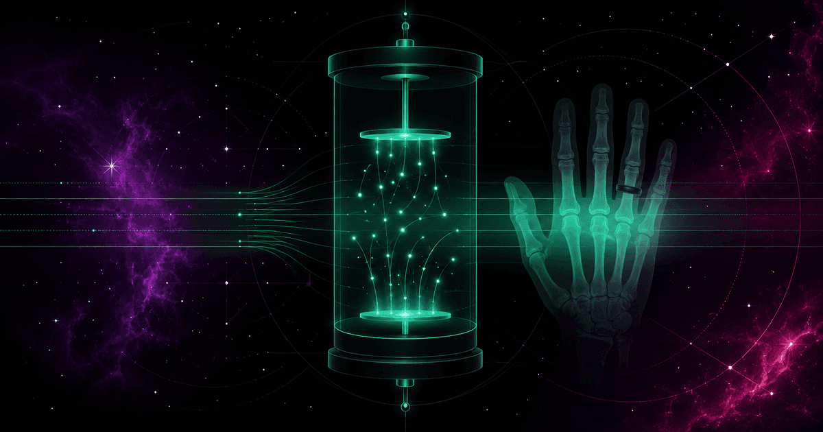
On the evening of Friday, 8 November 1895, at the Physics Institute of the University of Würzburg, Wilhelm Conrad Röntgen was running routine experiments on cathode rays. He had wrapped a glass Crookes tube tightly in black cardboard and darkened the room completely — yet he noticed a puzzling greenish glow on a nearby sheet coated with barium platinocyanide. An invisible force was passing through the cardboard, through thick books, through wood, and — what astonished him most — through the human body. Not knowing its nature, he named it the X rays, using the mathematical symbol for an unknown.
On 22 December 1895 Röntgen took the first X-ray photograph of a person. His model was his wife, Anna Bertha. Seeing the plate with the dark bones of her hand and the shadow of her ring, she is said to have cried, "I have seen my death." The discovery — awarded the first-ever Nobel Prize in Physics in 1901 — revolutionized medicine within weeks. But wonder came with an awkward question: how do you measure and put a number on something entirely invisible to the senses?
Why air, of all things
Ionizing radiation moves no needle and gives off no warmth to the touch — until it reacts with matter, it leaves no trace you can easily read. Physicists at the turn of the century needed a medium that was reproducible, universally available and homogeneous, in which the rays would produce a measurable effect. That medium turned out to be ordinary atmospheric air.
The choice was no accident. An X-ray or gamma photon passing through a gas collides with atoms of nitrogen, oxygen and argon and knocks electrons out of them. Ion pairs are formed: a free electron with a negative charge and a positive residual ion. That is ionization. If the process happens inside an ionization chamber — a metal vessel filled with air and holding two electrodes under voltage — the charges drift toward the electrodes of opposite sign, and their motion creates a faint saturation current directly proportional to the intensity of the radiation. The French physicist Jean Perrin described this principle as early as 1896, opening the era of gas dosimetry.
Air won for three reasons. A chamber filled with dry air is easy to build and faithfully replicate in any laboratory in the world — no rare or unstable reagents required. Air surrounds a person constantly, so it was a natural reference point. And, most importantly, the effective atomic number of air (Z_eff ≈ 7.6) is strikingly close to that of water (≈ 7.4) and soft tissue (≈ 7.4). That coincidence held out the hope that a measurement in gas would translate easily into what happens inside a patient's body.
The quantity describing this process was named exposure (denoted X): the total electric charge of one sign produced per unit mass of air. In the SI system its unit is the coulomb per kilogram (C/kg).
Anatomy of an "ugly" number
Before SI became standard, science ran on the CGS system — built on the centimetre, gram and second. It was in CGS, at the Second International Congress of Radiology in Stockholm in 1928, that the first international unit for a quantity of radiation was officially defined: the roentgen, symbol R. Its root was the Villard unit, proposed in 1908. The roentgen was the amount of X or gamma radiation that, in one cubic centimetre of dry air at normal conditions (0 °C, 1013.25 hPa), releases ionization equal to one electrostatic unit of charge (1 esu) of one sign.
The definition worked beautifully in the CGS era, but its conversion to SI startles the modern reader:
1 R = 2.58 × 10⁻⁴ C/kg
Where does such an un-round, seemingly arbitrary constant come from? It follows directly from the move from volume measures and electrostatic charges to mass measures and coulombs. Let us trace it step by step. One electrostatic unit of charge, in coulombs, is:
1 esu ≈ 3.33564 × 10⁻¹⁰ C
The density of dry air at normal conditions is ρ ≈ 1.293 kg/m³, so the mass of one cubic centimetre is:
m = 1.293 kg/m³ × 10⁻⁶ m³ = 1.293 × 10⁻⁶ kg
Exposure as the ratio of charge to mass therefore gives:
X = 3.33564 × 10⁻¹⁰ C ÷ 1.293 × 10⁻⁶ kg ≈ 2.5798 × 10⁻⁴ C/kg
When, in the second half of the 20th century, metrology set about tidying up non-SI units, the International Commission on Radiation Units and Measurements (ICRU) in 1971 rounded this value and adopted it as exactly 2.58 × 10⁻⁴ C/kg. That single number is a bridge between 19th-century physics of charges drifting in a droplet of gas and the modern metrology of kilograms and coulombs.
The four quantities forever confused
Exposure is just one link in a longer chain. In conversations about radiation, four different concepts are routinely thrown into one basket — yet each answers a different question and concerns a different stage: from the source, through space, to the effect in tissue.
| Quantity | SI unit | Old unit | Question it answers | Limitations |
|---|---|---|---|---|
| Activity (A) | becquerel (Bq) | curie; 1 Ci = 3.7 × 10¹⁰ Bq | How many decays occur in the source per second? | Source only; nothing about energy or direction |
| Exposure (X) | coulomb/kg (C/kg) | roentgen; 1 R = 2.58 × 10⁻⁴ C/kg | How much charge was produced in air? | X and gamma only, air only, up to 3 MeV |
| Absorbed dose (D) | gray (Gy) | rad; 1 rad = 0.01 Gy | How much energy did a kilogram of matter absorb? | Any radiation and any medium |
| Equivalent dose (H) | sievert (Sv) | rem; 1 rem = 0.01 Sv | How large is the risk to human health? | Living organisms only; accounts for biology |
In cause-and-effect terms it goes like this. Everything begins with activity — how often an unstable nucleus emits a particle or photon, whether or not anyone is nearby (we cover the becquerel in "The becquerel — the pulse of the microworld"). As the emitted photons cross space, they ionize the air they meet — and that ability is what exposure measures. When the radiation hits an obstacle, e.g. a patient's body, part of the energy is deposited in the tissue — that is absorbed dose in grays ("The gray that heals and kills"). But physics is not biology: the same dose harms differently depending on the type of particles and the sensitivity of the organ, so the real risk is captured only by equivalent dose in sieverts ("A banana, a flight and a CT scan").
The dark ages of dosing
An objective unit was introduced in 1928 not for academic reasons but in response to a tragic toll. For the first three decades after Röntgen's discovery, medicine operated in near-total metrological chaos, and ignorance of the biological effects bred recklessness.
In November 1896 the American physicist Elihu Thomson deliberately exposed the little finger of his left hand to the beam of an X-ray tube for tens of minutes a day. The result was painful swelling, blisters and stiff joints — and Thomson published one of the earliest warnings. Even so, it was long believed that the burns were caused not by the radiation but by electrostatic discharges around the tube. Only the careful experiments of William Rollins in 1898–1901, in which he placed guinea pigs in grounded Faraday cages, proved beyond doubt that the X-rays themselves destroy tissue.
Lacking physical units, radiologists had to measure radiation with... biology and chemistry:
- Skin erythema dose (SED) — the amount of radiation needed to produce, after a few days, a clear reddening of the patient's skin. The reaction depended on individual traits, and the effect appeared too late to prevent an overdose.
- Sabouraud–Noiré pastilles (1904) — discs of barium platinocyanide changed colour under the rays from green to orange-brown; the colour was compared against a chart. Judging it in the dim light of a consulting room was highly subjective.
- Holzknecht and Kienböck units (1902–1905) — based on chemical reactions of salts and on the degree of blackening of light-sensitive paper containing silver bromide.
A rising number of cancers and deaths among staff forced institutionalization. The British Roentgen Society issued the first protective recommendations in 1915, and in 1924 Arthur Mutscheller proposed the notion of a "tolerance dose" for workers — set at one tenth of the erythema dose per month. The Stockholm congress of 1928 finally established two bodies: the later International Commission on Radiological Protection (ICRP) and the ICRU. It was they who made the roentgen the first global unit.
The twilight of the roentgen
For all its merits, the roentgen had built-in limitations that eventually excluded it from modern metrology. It was measurable only for photon radiation (X and gamma) and only in dry air — it could not measure exposure from electrons (beta), protons, alpha particles or neutrons, all crucial in reactors and radiotherapy. Moreover, the definition broke down above 3 MeV: at such energies the secondary electrons knocked out of the air have ranges on the order of metres, and it becomes impossible to keep the whole charge inside the chamber, which rules out a precise measurement.
So physicists began to look for units based on energy rather than charge. In 1940 Louis Harold Gray proposed the "gram-roentgen", tying the measurement to the energy transferred to tissue. This led to the definition of the rad in 1953 (1 rad = 100 erg/g), and of the gray (Gy) in 1975, equal to the joule per kilogram. The final step was the sievert (Sv), introduced in 1979 as the unit of equivalent and effective dose in place of the old rem.
The energy bridge: the f-factor
How do you connect historical roentgens with the actual energy inside a patient's body? That is the job of the f-factor, and the relation is simple:
D = f × X
where D is the absorbed dose in a given medium, X is the exposure, and f depends on the chemical composition of the medium and on the photon energy. At the heart of the conversion is the mean energy needed to create one ion pair in air — about 34 eV, i.e. W/e ≈ 33.97 J/C. Looking at the values of f for different tissues shows why a measurement in air does not directly reflect the hazard to organs:
- Dry air — by definition f = 0.877 rad/R, which means an exposure of 1 R deposits 8.77 mGy in air.
- Soft tissue and muscle — thanks to a similar composition, f is stable at about 0.93–0.96 rad/R (1 R ≈ 9.3–9.6 mGy). Hence the old practical approximation: "one roentgen is roughly one rad in the body."
- Bone — has a higher density and a higher effective atomic number (Z_eff ≈ 13.8). At low energies (below 100 keV), typical of classic X-rays, photoelectric absorption dominates, and its probability grows as the cube of the atomic number. The f-factor for bone jumps sharply, reaching about 4.24 rad/R at 30 keV.
The conclusion is tangible: at an exposure of 1 R, soft tissue absorbs about 0.96 rad (9.6 mGy), but bone in the same time absorbs over 4 rad (40 mGy) — more than four times as much. Only above 1 MeV, where Compton scattering rules, does f for bone fall to 0.92–0.95 rad/R. These discrepancies prove that the roentgen cannot be a universal measure of patient dose.
Two myths to close on
"The roentgen tells you what a person received." It does not. The roentgen is a measure of exposure — it describes solely the ability of a stream of photons to ionize dry air under strictly defined conditions. As the f-factor shows, the same exposure gives a different dose in fat, muscle and bone, and on top of that the roentgen entirely ignores biology: it does not know that the thyroid or the marrow are far more sensitive than other tissues. The real dose and risk to a human are expressed only by the sievert.
"All radiation units say the same thing." Putting an equals sign between the becquerel, roentgen, gray and sievert is a category error. A source with an activity of billions of becquerels may pose no hazard at all if it sits inside a thick shield of concrete and lead. And conversely — a negligible absorbed dose in grays can carry an enormous risk (a high sievert value) if it reached a sensitive organ by inhaling alpha-emitting particles. Without accounting for physics and biology, these units are not directly interconvertible.
The evolution of radiation units is a story of gradually taming nuclear physics. The roentgen was the scaffolding on which modern radiological protection was raised — it let us abandon methods as dangerous as inducing controlled reddening on researchers' skin. Replacing it with the gray and sievert was not cosmetics but a deep reform: science separated the purely physical aspect (energy in matter) from the biological-medical one (risk to health). The roentgen remains a priceless relic — a memento of the days when physicists tried to capture an invisible force by counting subtle charges drifting in ordinary air.
Further reading
- Jerzy Sobkowski, Chemia radiacyjna i ochrona radiologiczna (Adamantan, 2009) — accessibly connects the physical chemistry of radiation–matter interaction with the practice of dosimetry and radiological protection.
- Andrzej Hrynkiewicz (ed.), Człowiek i promieniowanie jonizujące (Wydawnictwo Naukowe PWN, 2001) — a compendium on the effect of radiation on the body, with a clear account of how dosimetric units evolved.
- ICRU Report 33, Radiation Quantities and Units — the source definitions of exposure, kerma and absorbed dose.
- "Roentgen (unit)" entry on Wikipedia — a concise reconstruction of the unit's definition and conversions.