
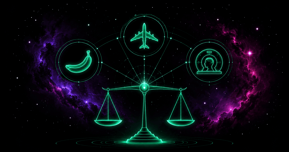
Picture a passenger at Warsaw's Chopin Airport eating a banana before a flight to New York. A few days earlier they had a three-dimensional dental scan in a cone-beam CT (CBCT). In a moment they will climb to ten kilometers, where the atmospheric shield is thinner and particles rain down from deep space. Three utterly different worlds: a fruit off a tree, a medical procedure from an X-ray tube, and a flight into the upper troposphere.
And yet each of these is described in the same unit — the microsievert (μSv). How is it possible that a banana, an airplane, and a CT scanner land on one scale? The answer lies in what the sievert really is. It does not measure the raw energy deposited in the body. The sievert is a biological translator: it converts the tangled physics of subatomic collisions into a single, statistical language of health risk.
The sievert is not the gray
In talk about radiation, four different quantities are constantly muddled together. They are worth separating:
- Source activity (becquerel, Bq) — the number of nuclear decays per second. It tells you how intensely a substance decays, nothing about how much radiation reached anything.
- Exposure (formerly the roentgen, R) — the ability of X-rays and gamma rays to ionize air. A historical measure of field intensity.
- Absorbed dose (gray, Gy) — the energy deposited in a kilogram of matter, 1 Gy = 1 J/kg. A purely physical, objective quantity — but, as we put it in a separate piece on the gray, biologically blind. It treats every deposited joule the same.
- Equivalent and effective dose (sievert, Sv) — the absorbed dose multiplied by factors that account for the harmfulness of a given radiation type and the sensitivity of specific organs.
The gray answers "how much energy?" The sievert answers "how dangerous is this to health?" In physical dimension both reduce to joules per kilogram, but their meaning sits at opposite poles: one measures the physical cause, the other the estimated biological effect.
Rolf Sievert and his portable chamber
The unit is named after Rolf Maximilian Sievert (1896–1966), the Swedish medical physicist who shaped the framework of modern radiation protection. Born in Stockholm into a wealthy industrialist family, he began with medicine at the Karolinska Institute, but physics turned out to be his true calling.
In 1920, seeing the chaotic growth of radiotherapy, Sievert offered the Radiumhemmet clinic his "voluntary and unpaid collaboration." There was a time when using radium and X-ray machines felt like groping in the dark: patients and staff suffered severe burns, and radiology's pioneers died of leukemia. Sievert grasped that the foundation of safety had to be the precise measurement of dose.
His most important invention, from 1926, was the condenser chamber — a miniature, portable dosimeter that worked without a permanent cable connection. The device was charged electrostatically, and passing radiation ionized the air inside and gradually discharged it; measuring the remaining voltage let you compute the dose. Thanks to the small spherical chambers, doctors could place them directly inside patients' body cavities, monitoring the dose reaching a tumor in real time. Sievert's 1932 doctoral thesis introduced, among other things, the so-called Sievert integral, describing the dose distribution around radium sources. Later he initiated Sweden's first national inspections of X-ray equipment and helped found the International Commission on Radiological Protection (ICRP), which he chaired from 1956 to 1962. Privately he was a passionate entomologist — he bequeathed his vast insect collection to the University of Lund.
How the sievert is built: two levels of weighting
Getting from objective energy (the gray) to risk (the sievert) takes two steps, defined by the ICRP.
Step one — equivalent dose (H). For a given tissue we multiply the absorbed dose by the dimensionless radiation weighting factor wᵣ, which reflects how severely a given radiation type damages cells — and we sum over all radiation types: H = Σ wᵣ × D.
The wᵣ values under the current ICRP Publication 103 (2007) recommendation:
| Radiation type | Factor wᵣ |
|---|---|
| Photons (X-ray, gamma), all energies | 1 |
| Electrons and muons (beta) | 1 |
| Protons and charged pions | 2 |
| Alpha particles, fission fragments, heavy ions | 20 |
| Neutrons | 2.5 – 20 (function of energy) |
For neutrons, wᵣ is not a constant but a continuous function of energy: from about 2.5 for thermal neutrons, through a peak of 20 near 1 MeV, down to about 5 for energies above 20 MeV.
Step two — effective dose (E). To assess whole-body risk under uneven irradiation, we sum the organs' equivalent doses, each multiplied by the tissue weighting factor wₜ — a measure of its susceptibility to radiation-induced cancer: E = Σ wₜ × Hₜ.
The wₜ factors under ICRP 103 (they sum to 1):
| Organs / tissues | Factor wₜ |
|---|---|
| Red bone marrow, colon, lung, stomach, breast | 0.12 each |
| Gonads | 0.08 |
| Bladder, liver, esophagus, thyroid | 0.04 each |
| Skin, bone surface, brain, salivary glands | 0.01 each |
| Remainder tissues (13 organs, averaged) | 0.12 |
A simple example closes the mechanism. If an examination delivers 10 mGy of gamma radiation (wᵣ = 1) to the thyroid alone, the thyroid's equivalent dose is 10 mSv. But the whole-body effective dose is only 10 mSv × 0.04 = 0.4 mSv — and only that number lets you compare such a scan with uniform whole-body irradiation. Effective dose is computed for an averaged "reference person"; it is a statistical tool, not a prognosis for an individual patient.
It is also worth remembering the old unit, the rem: 1 Sv = 100 rem, so 1 rem = 0.01 Sv — exactly as our equivalent dose converter works it out. Because the sievert is a very large unit, in everyday life we use milli- and microsieverts.
Why an alpha particle is twenty times more dangerous
The factor of 20 for alpha particles is not arbitrary — it follows directly from the microscopic geometry of life. The key is linear energy transfer (LET) and relative biological effectiveness (RBE).
The DNA double helix is about 2 nanometers across. X-rays and gamma rays are low-LET waves (below 10 keV/μm). A photon crossing a cell nucleus rarely deposits energy right next to the DNA strand, and if it does cause damage, it is most often an easily repaired single-strand break — the other strand then serves as a ready template.
An alpha particle (a helium nucleus: two protons and two neutrons) is massive, slow, and strongly charged. Its LET often exceeds 100 keV/μm. Crossing a cell, it leaves a dense, ionization-saturated track and, over a distance of just a few nanometers — exactly the scale of DNA's width — deposits a huge burst of energy at once. This tears both strands simultaneously at many neighboring points, creating complex double-strand breaks (DSBs). The cell triggers a molecular alarm — the ATM kinase phosphorylates thousands of H2AX histone molecules around the damage, forming γ-H2AX foci — but such intricate damage often cannot be repaired faithfully. The outcome is either apoptosis (cell suicide) or a faulty rejoining of strands that permanently damages tumor-suppressor genes and starts carcinogenesis. For this drastically higher efficiency at destroying the genome, alpha particles received the highest weighting factor.
The dose scale: from a banana to a reactor
Each of us continuously receives a dose from the natural background. In Poland, the National Atomic Energy Agency (PAA) assumes a default of about 2.4 mSv per year — the sum of cosmic radiation, the Earth's crust, isotopes in our bodies, and inhaled radon. The background varies wildly by geography, though: in Finland it averages around 7.3 mSv, and residents of Ramsar in Iran, where hot springs carry radium-226, receive over 260 mSv per year — and yet studies there show no elevated cancer incidence.
The table below places typical effective doses on one scale:
| Situation | Typical effective dose | Context |
|---|---|---|
| Eating a banana | 0.1 μSv | The "banana equivalent dose" — purely educational |
| Dental X-ray | 1–5 μSv | Local exposure of negligible risk |
| Chest X-ray | ~20 μSv | About 3 days of natural background |
| Warsaw–New York flight | 30–80 μSv | Secondary cosmic radiation at cruising altitude |
| Dental CT (CBCT) | 30–200 μSv | Depends on the field of view |
| Annual limit for the public | 1 mSv | Legal limit above background and medicine |
| Head CT | 1–2 mSv | Standard brain examination |
| Annual natural background in Poland | 2.4 mSv | PAA default value (with radon) |
| Average annual exposure of a Pole | 3.3–4.4 mSv | ~40% from medical diagnostics |
| Chest CT | ~7 mSv | Almost three years of natural background |
| Annual limit for a worker | 20 mSv | Standard for occupationally exposed staff |
| Average Chernobyl liquidator dose | 100–250 mSv | Emergency crews, 1986–1989 |
| Acute radiation syndrome threshold | ~500 mSv (0.5 Sv) | Onset of deterministic effects |
| Lethal dose LD₅₀/₃₀ (untreated) | 4–5 Sv | Death of half the exposed within 30 days |
Dose is not dose rate
The most common mistake in judging danger is ignoring time. You must always distinguish the cumulative dose (in Sv) from the dose rate (in Sv/h, mSv/h, or μSv/h).
Take today's zone around Chernobyl. In the city center the dose rate is about 0.15 μSv/h — a perfectly safe value, close to the background in many parts of Poland; a whole day spent there delivers less than a short flight. Now the day of the 1986 disaster, when rescuers worked next to the wrecked reactor, where the dose rate exceeded a dozen or more sieverts per hour: it took only a quarter of an hour to receive a dose of several sieverts.
The difference is biologically fundamental. The body has an efficient DNA-repair system. A dose of 100 mSv spread over 50 years lets cells repair the rare breaks as they occur, and the statistical rise in risk is nearly undetectable. The same dose delivered in a few seconds can overwhelm the repair machinery and fix mutations in place. That is why radiation protection uses the DDREF factor, which lowers the estimated risk for slowly received doses.
Three dosimetric traps
"The gray and the sievert are the same thing, since both are joules per kilogram." Mathematically, yes, the dimension is identical — but the meaning is entirely different. The gray measures the physical cause, the sievert the estimated effect. If the lungs absorb 0.1 Gy from alpha-emitting radon, the equivalent dose is as much as 2 Sv; the same 0.1 Gy of gamma radiation gives 0.1 Sv. An identical energy deposit, a twentyfold difference in harm.
"Every dose, however tiny, carries lethal risk." This is a literal reading of the linear no-threshold model (LNT), formulated on the basis of Hermann Muller's fruit-fly studies from the 1920s (popularized in his 1946 Nobel lecture). LNT is a convenient and safe assumption for designing standards and shielding, but at doses below ~100 mSv there is no direct epidemiological evidence of an increase in cancer. We evolved in a constantly radioactive environment and have efficient DNA-repair mechanisms. UNSCEAR explicitly warns against multiplying minimal background doses by millions of people to count "virtual victims."
"A CT scan is either a trivial routine or a mortal danger." The truth is in between. A chest CT deposits about 7 mSv — nearly three years of background — so it should not be performed without a clear justification. On the other hand, scaring patients with death is an abuse: the statistical risk of a fatal cancer from a 10 mSv dose is about 0.05%. In urgent diagnostics (an accident, a suspected aneurysm or tumor), the benefit of a fast diagnosis vastly outweighs the minimal, time-delayed risk. Doubts are always worth discussing with the attending physician or a medical physicist.
The banana, potassium, and homeostasis
Let us return to our banana. The concept of the banana equivalent dose (BED) became popular as proof that natural food, too, can be radioactive. That is true: bananas are rich in potassium, and natural potassium contains about 0.012% of the radioactive isotope potassium-40. Eating a fruit gives, in theory, about 0.1 μSv.
As an educational tool for taming the fear of radiation, BED works beautifully — but it hides a methodological error. The body maintains very strict potassium homeostasis: the kidneys police its level, and any excess is quickly excreted. Eating a banana therefore does not permanently raise the amount of potassium in the body, or our internal radioactivity. Regardless of diet, we carry natural potassium-40 generating thousands of decays per second (an activity of about 4–5 kBq). The banana does not change that — it merely adds, briefly, to a pool we are about to level out anyway.
A compass of risk, not a verdict
The sievert is one of the most successful ideas in the history of medical physics. It lets us reduce subatomic phenomena — from high-energy cosmic protons striking an aircraft's fuselage to low-energy photons from an X-ray tube — to a single, readable number describing biological risk. That is why a banana, a flight, and a CT scan can be placed on a common axis at all.
You only have to remember that the effective dose in sieverts is a statistical measure, created to protect populations, not a clinical prognosis for a particular person. A few millisieverts from a CT scan are not a verdict but a small, estimable shift in probability over an entire lifetime. Thanks to the sievert we have a precise compass that lets us function rationally in a world where radiation is the natural background of our existence.
Further reading
- J. Sobkowski, M. Jelińska-Kazimierczuk, Nuclear Chemistry (Adamantan, Warsaw 2006) — the basics of dosimetric quantities and units.
- B. Gostkowska, Radiation Protection. Quantities, Units and Calculations (Central Laboratory for Radiological Protection, Warsaw 2018).
- ICRP Publication 103 — The 2007 Recommendations of the International Commission on Radiological Protection, Annals of the ICRP 37(2–4), 2007: the source of the wᵣ and wₜ factors.
- UNSCEAR, reports of the UN Scientific Committee on the Effects of Atomic Radiation — data on background doses and the health effects of low doses.